Laparoscopic cholecystectomy
|
Laparoscopic Cholecystectomy involves introducing 4 'ports' into the abdomen by making tiny cuts - two cuts are 1 cm in length each, and the other 2 are only 0.5 cm long. Ports are special cannulas or tubes, through which the camera & intruments are passed into the abdomen. To create space, the abdomen is filled up with CO2 gas, and the ports have valves that prevent gas-leak when passing intruments through them.
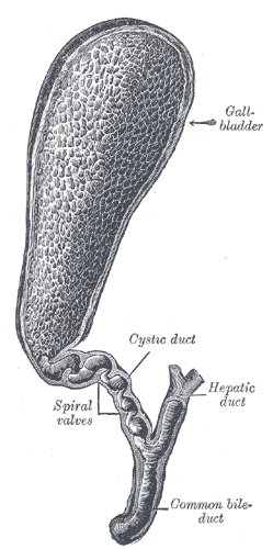
The gall bladder is connected to the common bile duct by a small tube called the cystic duct. This cystic duct is clipped off & divided, as is the blood vessel supplying the gall bladder. This allows the gall bladder to be detatched from the underside of the liver. The free gall bladder is put into a retrieval bag & pulled out from the cut at the umbilicus.
In some cases, it becomes advisable to carry out an x-ray examination of the common bile duct, to check if any gall stones have escaped out of the gall bladder into the bile duct. This examination is called a 'peroperative cholangiogram'.
The patient receives a general anaesthetic during the operation, and in most cases is able to leave the hospital on the same evening or the following morning.
|
|
|
|
| |